You must be signed in to read the rest of this article.
Registration on CDEWorld is free. You may also login to CDEWorld with your DentalAegis.com account.
This case study exemplifies how consistent communication between the dental laboratory and practitioner can yield a workable treatment plan that best suits the patient’s goals, streamlines the case processing, and provides a finished product that is both optimally functional and esthetically pleasing.
Case Study
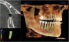
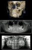
A middle-aged female presented with moderate-to-severe vertical bone loss, generalized mobility (Class II), and generalized probing depths of 6 mm or more with bleeding and/or exudate on all probings (Figure 1). The patient and dentist agreed to a treatment plan that involved the removal of all teeth and the fabrication of full upper and lower immediate dentures with the possibility of implant-retained appliances at a later date.
Records were taken along with a face bow so that the orientation of the dentition could be accurately transferred when the laboratory mounted the case on a semi-adjustable articulator. In addition to discussing the treatment plan with the laboratory, the dentist provided detailed pre-operative pictures, which significantly aided in the subsequent smile design. These photographs revealed the position of tooth No. 8 to be acceptable and tooth No. 9 to be supererupted far below the smile line and facially protruded. In order to capture the anterior information on the mounted models, the laboratory marked the line at the bottom of tooth No. 8 in red and shaved tooth No. 9 even with tooth No. 8 so that the team could achieve an even curvature of the smile line. Then the buccal portion of tooth No. 9 was marked in red and shaved back to become even with tooth No. 8 in order to obtain harmony in the arch form. Then, the laboratory fabricated an anterior rubber index to capture the orientation of the adjusted upper anteriors.
Prior to extracting the teeth from the upper model, the desired midline was marked in red. The cervical of the buccal and lingual of all the upper teeth was also traced in red. Next, all the upper teeth were extracted from the model using a long, tapered crosscut carbide bur attached to a handpiece. Only the clinical crown was removed, with no socketing or depression at the site of each tooth. The buccal portion of the extraction sites and the papilla areas were smoothed with the bur to approximately 1 mm to obtain a rounded, smooth ridge. Next, the upper anterior teeth were set using the rubber index as a guide.
Veracia SA (Shofu, www.shofu.com) semi-anatomical teeth in shade A2 were chosen because they are innovative in composition, consisting of a micro-filled hybrid composite reinforced with layered glass to give the teeth superb esthetics, extended durability, and exceptional bonding strength with the denture base. The tooth material is also homogenous, eliminating concern about fracturing of layers (Figure 2).
After the upper anteriors were set to the rubber index, the lower teeth were removed from the model following the same technique. Then, the lower anterior teeth were set onto the ridge, trying to come as close to an ideal setup as possible. Interoral measurements during diagnosis in relation to overjet and overbite were considered.
The retromolar pad on both sides of the lower posterior was circled, and a line was drawn horizontally two-thirds up the retromolar pad. Teeth should not exceed this line. A vertical line was also drawn from the center of the retromolar pad on both sides over the center of the ridge to the distal of the lower cuspid area. The central fossae of each lower posterior tooth was set directly over the line on the center of the ridge to maximize functionality and stability of the lower arch. Then the upper posteriors were placed, checking working and balancing movements after setting each tooth.
The upper and lower arches were waxed using Dimensions Gingival Wax (Hi-Tech Dental Supply, www.hi-tecdental.com), which affords the patient a more realistic preview of how the finished dentures might look after processing and naturalization (Figure 3). The case was sent to the dentist for verification. After the dentist and patient approved the setup, the case was returned to the laboratory for processing.
After the case was finished, it was naturalized with Ceramage Gum Color Kit (Shofu), which was designed to allow the technician to esthetically enhance the denture base with natural gum colors. The Ceramage Gum Color Kit is unique on the market because of its 73% zirconia content, which adds strength and luster. Ceraresin Bond (Shofu) was used as the bonding agent.
Because this was an immediate upper and lower case, the laboratory fabricated silicone models of the inside of each finished and polished appliance. On these models, the laboratory fabricated clear surgical templates as a guide for the dentist during insertion of the dentures after extractions
Conclusion
Because the dentist took considerable care at the impression stage, rechecked his records, properly utilized the face bow, and provided numerous pre-operative pictures, the laboratory was efficiently able to fabricate appliances that satisfied the patient’s esthetic and functional needs with minimal adjustments (Figure 4).
Disclosure
This article was supplied by Shofu Dental Corporation.
Take the quiz online to receive CE credit: dentalaegis.com/go/idt718
About the Authors
James Collis, CDT
James Collis, CDT, has been a certified dental technician and an actively practicing laboratory technician for more than 30 years. He possesses a degree in dental technology from Triton College. His laboratory, Collis Prosthodontic Laboratory, specializes in high-quality, removable prosthodontic appliances in suburban Chicago. Mr. Collis previously served as an instructor in the junior/senior laboratory of Northwestern University Dental School for 10 years. In that capacity, he presented numerous courses on prosthetics, including advanced esthetics pertaining to removable prosthodontics, fixed work, and advanced attachment and implant techniques. Mr. Collis has been active on the lecture circuit for the past 10 years. He is an advisory board member for Spectrum Dialogue and has written numerous articles on a wide variety of topics in industry publications.
Neeraj Khanna, BSc, DDS
Neeraj Khanna, BSc, DDS, completed his bachelor of science degree at the University of Toronto in Canada and received his DDS degree in 1993 from the University of Detroit Mercy-School of Dentistry. In 1994, Dr. Khanna completed a general practice residency at the University of Rochester-Strong Memorial Hospital in Rochester, New York. After being an associate dentist for several years, Dr. Khanna opened his practice in 2000 in Geneva, Illinois. Dr. Khanna also completed training at the prestigious Dawson Academy in St. Petersburg, Florida, and has been involved as an associate faculty member since 2010. Dr. Khanna is now president of the Khanna Institute of Dental Excellence and has lectured in the United States, India, and United Arab Emirates.